What is so fascinating or even better said the beauty of working with tiny worms? Many might be curious about this, when they meet researchers like me. I would suggest to look at the marvelous wonders of the hairybellies, the phylum Gastrotricha, and you will understand why it is an amazing world you are entering when working with these tiny worms of the meiofauna. Meiofauna in general are animals, which will pass through a sieve of a size of 500 µm, only half a millimeter. They usually live in the space between the sand grains that is the interstitium. This means they do not move the sand grains, but glide through the small crevices between the different-sized sand grains. Imagine this; these animals can easily squeeze themselves through these tiny spaces and pores, which we cannot even see with our naked eyes. We need at least a magnifier for seeing the pores.
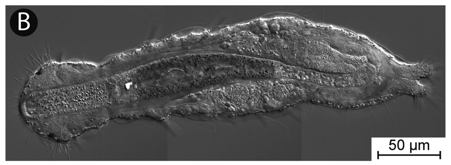
About 760 species are known to date occurring both in freshwater and marine habitats. The first gastrotrich was described from freshwater and afterwards they thought be exclusively freshwater. However, with the recognition of the interstitial realm in the middle of the 20th century many new marine species have been discovered. Since then, it has become apparent that indeed the majority of species is actually marine. As written above, the animals are tiny with body length of just 0.1 mm to a maximum of 1 mm. When one extracts them from their habitat to investigate them further, one often can see unicellular protists like ciliates alongside them. These protists often pass them by and can be three to four times larger than the gastrotrich. Bear in mind, we are speaking of a single cell being that much larger than an organisms with hundreds to thousands of cells.
How do we find these animals, when they are so small that we cannot see them with our naked eyes? We usually use a method that helps us to concentrate the specimens from a habitat sample and then sort this concentrated sample under a dissecting microscope using magnifications of at least 20fold, but often 40 to 64fold. This degree of magnification is only sufficient for finding them. For species identification, higher magnifications of up to 1000x are needed and in a few cases even this is not enough and only electron microscopy can help.
How do we concentrate the specimens? I will present you here one method used in the study of the marine interstitium. The sediment sample is treated with a magnesium chloride solution, which has the same osmolarity as the seawater from which the sample was taken (it is usually 8% weight per volume). The magnesium ions replace the calcium ions in the cells and hence relax the specimens. This means they cannot attach themselves to, for example, sand grains. Then the sample is gently, but thoroughly stirred up and poured through a sieve with a mesh size of 64 µm. As the sand grains have a higher density than the specimens, the former will settle much faster than the latter and hence they get separated from each other in this process. In theory, “only” the specimens will end up in the sieve. As you can image this is not a failure-proof method and a lot of other stuff comes also along with the specimens, but it still concentrates the specimens and makes sorting much faster.
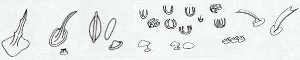
Even though gastrotrich are so small, they show an amazing array of delicate structures in their skins. As you can see on the figure below, they have different scales and spines in the so-called endocuticula of their skin. Their skin is composed of three parts. Like, for example, insects and earthworms they have a protective cuticula on their epidermis, which consists of a thin exocuticula and a thick, fibrous endocuticula. The scales and spines are embedded in the endocuticula, but the underlying epidermis builds them. Interestingly, in some gastrotrichs, this epidermis is syncytial. This means all epidermal cells have merged to one large cell with several nuclei. A similar process can also be found in our muscle fibers. Another interesting aspect about the scales and spines is that the most elaborate and largest (in relation to body size) ones can be found in the smaller gastrotrichs. This could indicate that like in small pelagic organisms these structures have a protective function. However, our knowledge about their function, their development and their genetic basis is very limited.
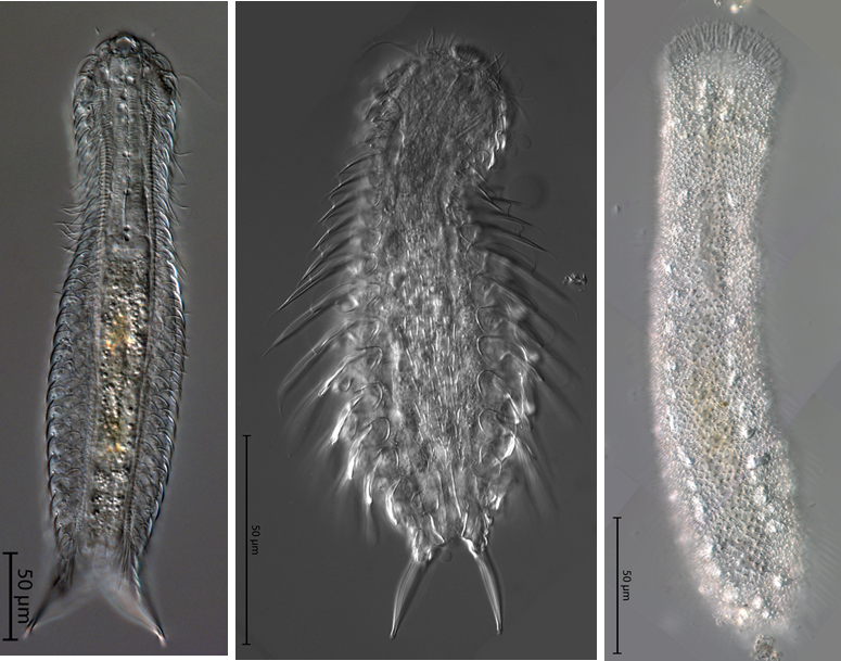
Gastrotrichs move by ciliary movement. They have cilia on their belly, which are used to push the animal forward; hence, both the English and Latin names mean cilia on the belly. An anecdote is that the first English common name, hairybacks, for them was actually as misnomer. The cilia of gastrotrich are unique across the tree of life. When organisms have cilia, these are not covered by the cuticula if they have one. If the cuticula gets very thick like in insects or crustaceans, then the cilia are lost. However, in gastrotrichs this is different. Here the cilia are covered by the thin exocuticula. This is a unique feature of the group. Why they have this feature, we do not know. For example, flatworms and acoels also move by ciliary movement and the cilia are not covered by something like a cuticula or parts of it.
Another interesting, but somewhat annoying feature of gastrotrichs is that they have a duo-gland system to attach themselves to a surface very quickly. A duo-gland system is a gland, which contains two types of gland cells. In this case, one of the glands releases a glue, which allows an extremely rapid attachment to a surface, usually the nearest sand grain. The other gland allows then a rapid release of animal from the surface again if needed. The glue is very effective and can withstand very strong shear and pull forces without detaching the animal from the substrate. The advantage of this system for the animal is that when the habitat is heavily stirred up, for example, due to a storm, the animal attaches itself to a sand grain. When the sand grain then settles again after the stirrup, the chances are high that this will happen in a habitat, which is also appropriate for the animal itself. These gland systems are not only known from gastrotrichs, but also other interstitial animals, but gastrotrichs are often especially sticky. These glands can be found at the feet at the posterior end of the animals and in several species at different parts of the body such as the adhesive tubes, which can often be seen to stick out from the body surface. Why is this cool feature annoying? Well, when one wants to transfer a gastrotrich from one dish to other using a pipette, one stirs up the water while sucking it in and then when one does not work very quickly the animals stick themselves to the inner surface of the pipette. As they are very good at sticking themselves to surfaces it can take quite a while and effort to get them out of the pipette again and sometimes they are lost forever.
Despite being so tiny, one can find almost all organs present in larger animals. They have a brain with a nervous system. They have different sensory organs including sometimes eyes. They have a kind of kidney. They have muscles. They have a gut. However, they lack a heart, gills and blood. They are so tiny and flat that they just do not need them. All components can be easily directly distributed in the body without the necessity of a transport system. Oxygen can be taken up directly from the environment through the skin as they have an excellent surface to volume ratio – lots of surface for very little volume.
We are interested in them as part of our InvertOmics, ERGA and BGE projects. We want to sequence the genomes of different species to get some clues on our knowledge gaps outlined above as well as to decipher their phylogenetic relationship. It is still not clear to which other animal groups they are most closely related; except for that they are part of a group comprising half of all animal phyla, also known as Lophotrochozoa or Spiralia. However, sequencing their genomes is a challenge by itself. As they are so tiny, one cannot get much DNA out a single specimen. However, working with single specimens is a huge advantage when sequencing reference genomes. Together with Nick Roberts and Kevin Kocot of the University of Alabama in the US, we are therefore working on protocols to get the most out of very little DNA from a single specimen. We made some progress and Nick is presenting first results on it this very week at the Biodiversity Genomics Conference 2022. His talk will be on Friday, October 7th, in the session on “Genome Project Symposia” from 1 pm to 3 pm BST if you want to get a sneak peek.
![]()