When an organism dies, it decomposes, whereby complex structures are broken down. Broadly following 5 to 6 stages, decomposition may be halted or accelerated at any point depending on conditions, such as temperature, water, and oxygen, resulting in different morphological and chemical changes to the organism. By chance, remains can be amazingly preserved in nature (unintentionally), resulting in fascinating fossils across geological time and surprisingly life-like humans found in peat bogs (Fig. 1) (Pestka et al., 2010). But we cannot solely rely on naturally preserved remains, due to the unpredictability and physical alterations of these processes. How, then, can we more precisely and systematically preserve tissues and morphologies?

Methods of fixing (preserving) organisms and tissues for scientific study are often specific and temporally unique. Historically, tissue fixation has focused on hardening the specimen to halt decomposition, allowing for thin sectioning (cutting) to be conducted more easily for histological assessments (studying tissue types and cell structures). The precursor to modern day scientific fixation began in the 1660s with boiling, followed by alcohol preservation and the injection of mounting agents in the 1700’s. In 1833 the first use of a “hardening agent”, chromic acid, was recorded (Jones 2001). Over time, various acids and chemicals were used as fixatives, in many unique historical preservation methods. However, during the turn of the 20th century, one chemical method, formalin, began to be commonly implemented (Jones 2001).
Namely, formalin is the diluted form of the organic compound formaldehyde (H2CO). To this day, in addition to being used for specimen fixation, formaldehyde is commercially used for industrial polymer production or biocides (Seymour and Kauffman, 1992). It was first synthetically produced for commercial use by Aleksandr Mikhailovich Butlerov in 1858. Formalin was first used as a scientific fixative by Dr. F. Blum in 1896, who considered it for this application when he noticed that it made the skin on his fingers stiffen (Seymour and Kauffman, 1992; Jones, 2001). Since then, formalin has been regularly used as a tissue preservative for natural history museums and scientific investigations.
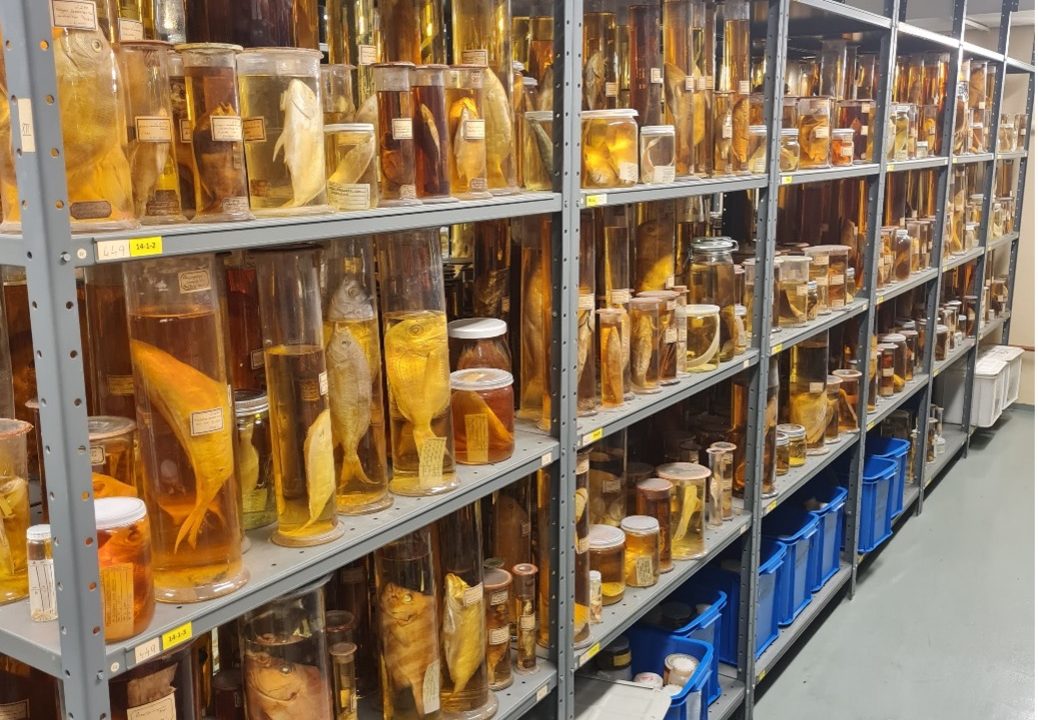
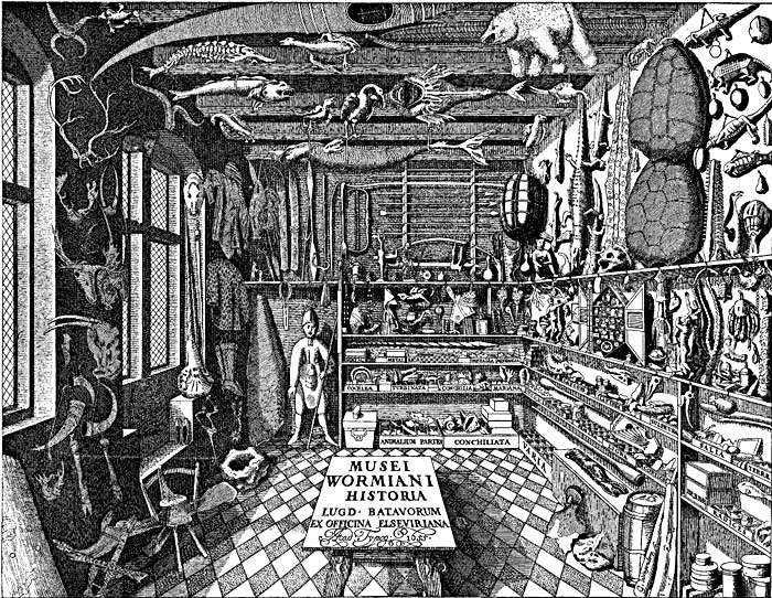
Collecting and preserving specimens for both private and public collections underwent a surge of popularity in the 18th and 19th century, owing to increases in global trade, communication, and travel, alongside the general popularity of natural history collections (e.g., cabinets of curiosities; Fig. 2) (Coote et al., 2017). Historic collectors, often-times traipsing through rural and/or dangerous environments, would frequently use formalin, due to its lower cost and convenience as opposed to alcohol. This was owing to the fact that formalin could be concentrated (reducing the initial volume of necessary liquid at departure), stores well long term, is incredibly versatile, and functions well in many conditions and across a range of tissue types (Fox et al., 1985). Unfortunately, drawbacks to formalin include tissue shrinkage, depending on the concentration used, and inherent health concerns, such as severe allergies, lung damage, kidney damage, and various cancers when continually exposed (Fischer, 1905; Musiał et al., 2016). Additionally, formalin preserves specimens by stiffening tissue(s) into which it is injected or soaked; stiffening chemically occurs through the random crosslinking of proteins together, and proteins to DNA, resulting in molecularly dense and unstructured tangled masses. This proves to be a major hurdle for modern sequencing endeavours, especially when unbuffered formalin is used. Many molecular technologies rely on DNA to be in a linear state to be sequenced, but formalin tangling obscures sequencing signals. Historically, the quantity of formalin used has been poorly documented for museum specimens, leaving us to guess quantities and preservation methodologies, with the added understanding that the longer specimens are exposed to formalin, the more molecular damage that occurs (Campos and Gilbert, 2012).
Approaches and protocols are actively being developed to reverse or repair formalin damage, such as those provided by Gould et al. (2021). These procedures are still being refined for specific tissue types and species, and the perfect solution has not yet been found. However, we remain optimistic and open to trying these procedures and tweaking them to be able to sequence formalin damaged crosslinked DNA. With such protocols, scientists, especially within Natural History Museums, could unlock information hidden in historic, formalin fixed specimens.
References:
Campos, P.F. and Gilbert, T.M.P. (2012) ‘DNA Extraction from Formalin-Fixed Material’, in Shapiro, B. and Hofreiter, M. (eds) Ancient DNA: Methods and Protocols. Totowa, NJ: Humana Press (Methods in Molecular Biology), pp. 81–85. doi:10.1007/978-1-61779-516-9_11.
Coote, A. et al. (2017) ‘When commerce, science, and leisure collaborated: the nineteenth-century global trade boom in natural history collections’, Journal of Global History, 12(3), pp. 319–339. doi:10.1017/S1740022817000171.
Fischer, M.H. (1905) ‘THE TOXIC EFFECTS OF FORMALDEHYDE AND FORMALIN’, The Journal of Experimental Medicine, 6(4–6), pp. 487–518.
Fox, C.H. et al. (1985) ‘Formaldehyde fixation.’, Journal of Histochemistry & Cytochemistry, 33(8), pp. 845–853. doi:10.1177/33.8.3894502.
Gould, A. L., Fritts-Penniman, A., & Gaisiner, A. (2021). Museum genomics illuminate the high specificity of a bioluminescent symbiosis for a genus of reef fish. Frontiers in ecology and evolution, 9, 630207.
Jones, M.L. (2001) ‘To Fix, To Harden, To Preserve–Fixation: A Brief History’, Journal of Histotechnology, 24(3), pp. 155–162. doi:10.1179/his.2001.24.3.155.
Musiał, A. et al. (2016) ‘Formalin use in anatomical and histological science in the 19th and 20th centuries’, Folia Medica Cracoviensia, 56(3), pp. 31–40.
Seymour, R.B. and Kauffman, G.B. (1992) ‘Formaldehyde: A simple compound with many uses’, Journal of Chemical Education, 69(6), p. 457. doi:10.1021/ed069p457.
![]()